Strong dental planning starts long before a drill touches a tooth. It begins with clear images that show what your eyes cannot see. Modern imaging tools let your dentist measure bone, spot hidden decay, and plan safe treatment. These tools guide simple fillings, complex root canals, and NE Philly dental implants. They also help prevent surprises during surgery. As a patient, you deserve to know what is happening inside your mouth. Clear pictures support honest talks about choices, costs, and risks. They also help your dentist match your goals with what is possible. First you see the problem. Then you agree on a plan. Finally you move forward with fewer doubts. This blog explains how common imaging tools work, why they matter, and what you should expect during a visit. You will see how better images lead to steadier results and stronger long term oral health.
Why dental imaging matters for you
You only see teeth and gums in the mirror. Your dentist must see roots, nerves, and bone. Imaging tools give that view. They turn guesswork into clear facts. They help your dentist:
- Find decay between teeth before pain starts
- Check bone height and thickness for implants
- Plan safe paths around nerves and sinuses
The National Institute of Dental and Craniofacial Research explains that early detection cuts tooth loss and pain from decay. Clear images support that early action.
Common dental imaging tools you may see
Your dentist may use more than one type of image. Each tool shows a different part of your mouth. Each one supports a different step in your plan.
Bitewing and periapical X rays
Bitewings show the crowns of your back teeth. They reveal decay between teeth and bone loss from gum disease. Periapical images show the whole tooth from crown to root tip. They help with root canals and cracked teeth.
Panoramic images
A panoramic image wraps one wide picture around your jaws. It shows all teeth, jaw joints, sinuses, and jawbone. It helps spot cysts, impacted teeth, and some tumors. It also gives a first look at bone levels for implants.
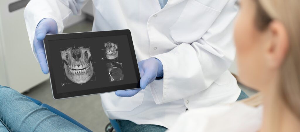
Cone beam CT scans
Cone beam computed tomography, or CBCT, creates a 3D view. It lets your dentist measure bone depth and width with high precision. This tool is useful for implants and complex extractions. It also helps avoid nerves and other weak spots.
Intraoral cameras
An intraoral camera is a small wand with a light. It sends close-up pictures of your teeth and gums to a screen. You can see cracks, stains, and worn fillings. These photos help you understand why a treatment is needed.
Comparing common dental imaging tools
| Imaging tool | What it shows best | Common use | Visit comfort
|
|---|---|---|---|
| Bitewing X ray | Decay between back teeth | Check for cavities and bone loss | Small sensor. Brief bite |
| Periapical X ray | Whole tooth and root tip | Root canals and tooth pain | Sensor near tooth. Short hold |
| Panoramic image | Both jaws in one view | Wisdom teeth and jaw review | Stand still. Machine moves around the head |
| Cone beam CT | 3D bone and nerve paths | Implant and surgical planning | Sit or stand. Brief scan |
| Intraoral camera | Surface cracks and wear | Patient education and records | Small wand. No bite needed |
Radiation, safety, and your peace of mind
Many people fear X-rays. You may worry about radiation. That concern is natural. Modern dental imaging uses very low doses. Digital sensors cut exposure compared with older film methods.
The US Food and Drug Administration gives clear guidance on dental X-ray safety. You can use that information to ask focused questions about your own care.
Your dentist should also:
- Use lead aprons and thyroid collars when needed
- Order images only when they change your care
- Review older images before taking new ones
How imaging shapes your personal treatment plan
Imaging is not just a picture. It is a map. It guides three key steps in your plan.
Step 1. Detect problems early
X-rays and photos reveal decay and bone loss before you feel pain. Your dentist can treat a small cavity with a simple filling instead of a root canal. This protects tooth strength and lowers cost.
Step 2. Plan safe and steady treatment
For NE Philly dental implants or other complex care, imaging shows if you have enough bone. It helps pick the implant size and angle. It also shows nerves and sinuses that need protection. That planning lowers the risk of numbness and sinus trouble.
Step 3. Check results and track change
After treatment, follow-up images confirm healing. They also track bone levels and watch for new decay. Your dentist compares images over time. This helps spot patterns like grinding or gum disease that may need new steps.
What you should expect during imaging
Most dental imaging is quick and free of pain. You can expect three simple stages.
- Set up. A staff person places a sensor or film in your mouth or has you stand in a machine. You may wear a lead apron.
- Position. You bite gently or stay still for a few seconds. The staff person steps away to take the image.
- Review. Images show up on a screen. Your dentist can zoom in and point to key spots.
If you gag easily or feel fear, speak up early. Your dentist can use smaller sensors, adjust angles, or take breaks. That respect for your comfort should be part of your care.
How to use images to ask stronger questions
Images help you take an active role. When you see your own teeth on a screen, you can ask direct questions.
- What does that dark spot mean for this tooth
- Can you show me the nerve and the implant on this scan
- What happens if I wait six months before treating this
Honest answers build trust. They help you weigh trade-offs between cost, time, and risk. That shared review can ease fear for both adults and children.
Key takeaways for your next visit
Imaging tools are not extras. They are core parts of sound dental planning. They:
- Reveal hidden problems before pain starts
- Guide safe treatment for fillings, root canals, and implants
- Support clear talks about options and long-term goals
At your next visit, ask which imaging tools your dentist plans to use and why. Ask to see the images. Ask what they show about your future oral health. You deserve clear pictures and clear answers.